Since the discovery of X-rays, fundamental physics has been a source of ideas for radiography and medical imaging. A new imaging method firmly rooted in particle physics was chosen by Time magazine as one of its “Inventions of the Year 2000”.

These days, innovation is flourishing in every industry. The variety of ideas currently being generated was illustrated last year by the news magazine Time, when it selected three specialist areas – consumer technology, medical science, and basic industry – in which to put new developments to the vote as “Inventions of the Year”.
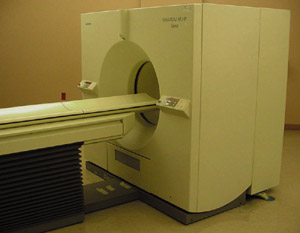
The award-winning invention in the medical science category was a scanner that combined the advantages of computer tomography with positron emission tomography (PET). The use of these techniques, which depend on detecting and analysing electromagnetic radiation (X-rays or gamma rays respectively), show that detection techniques from particle physics have made, and continue to make, essential contributions to medical science.
Tracers and tomography
Soon after their discovery by Roentgen in 1895, X-rays were being used for monitoring bones, teeth and other dense organic matter, thereby revolutionizing medical diagnostics and introducing a new science – radiography.
Then came nuclear medicine. George de Hevesy was awarded the 1943 Nobel Prize for Chemistry for his invention of radioactive tracers, in which small doses of radioactive material are administered to patients to follow the metabolic functioning of organs such as the kidneys or thyroid gland. The impact of the technique was so great that the supply of suitable radioactive isotopes went on to become an industry in its own right.
Although they provided valuable new information, these techniques, like conventional X-ray photographs, could only reveal a two-dimensional image of a three-dimensional body, and interpretation could therefore be difficult.
The imaging capabilities of X-rays were dramatically boosted by the 1972 invention of the computer-assisted tomography (CT) scanner, in which a fan-like beam of X-rays rotates round a patient, providing a two dimensional picture of a “slice” of their body. A complete three-dimensional image can be built up by scanning the body slice by slice. Tomography can be combined with nuclear medicine, for example in single-photon emission computed tomography (SPECT), which maps the internal distribution of the tracer.
The birth of PET
Many artificial isotopes emit positrons, the antiparticles of electrons. In the early 1950s it was discovered that these isotopes offered new possibilities for nuclear medicine.
A positron, once it has been produced, is quickly snapped up by a neighbouring matter particle – usually an electron. This annihilation of the positron and the electron produces a characteristic fingerprint – two 511 keV photons (gamma rays) shooting out in exactly opposite directions. By picking up these pairs it is possible to pinpoint where the positron annihilations occurred. The new science of PET was born, in which the annihilation signals track a patient’s metabolism, revealing, for example, the way in which the brain reacts to stimuli.
Since its inception, PET technology has profited from new developments in radiation detection, first using sodium iodide crystals, then using materials such as bismuth germanate (BGO), which offered better performance, and more recently lutetium oxyorthosilicate, which is faster and gives more light output than BGO.
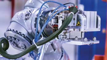
Specialists from particle physics have also applied new high-speed computing solutions (transputers) to speed up the imaging process.
Dedication’s what you need
Developing these techniques is time-consuming and requires a great deal of motivation as well as resources. In a pure science laboratory like CERN, which focuses on major experiments in particle physics, such spin-off research and development work has sometimes had to take a back seat, and several enthusiasts have left for new pastures.
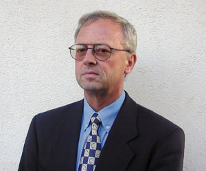
Among them is physicist David Townsend, who worked at CERN in 1970-1978 before concentrating on PET developments in the US and Europe and finally transferring to the University of Pittsburgh. He is one of the masterminds behind the development honoured by Time. The other is engineer Ronald Nutt, senior vice president and director of research and development at CTI PET Systems – the leading supplier of PET technology – in Knoxville, Tennessee.
Another physicist, Alan Jeavons, who worked at CERN with Townsend in the 1980s to develop the high-density avalanche chamber (HIDAC) PET camera, left physics research to form his own company, Oxford Positron Systems, and he has landed his first major contract to supply advanced PET systems.