Advanced scintillating materials have found their way into novel detectors for physics and medicine.

Image credit: M Hamonet (CPPM) & Luc Bidaut (Lincoln Univ. UK).
The Crystal Clear (CC) collaboration was approved by CERN’s Detector Research and Development Committee in April 1991 as experiment RD18. Its objective was to develop new inorganic scintillators that would be suitable for electromagnetic calorimeters in future LHC detectors. The main goal was to find dense and radiation-hard scintillating material with a fast light emission that can be produced in large quantities. This challenge required a large multidisciplinary effort involving world experts in different aspects of material sciences – including crystallography, solid-state physics, luminescence and defects in solids.
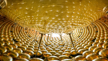
From 1991 to 1994, the CC collaboration carried out intensive studies to identify the most adequate scintillator material for the LHC experiments. Three candidates were identified and extensively studied: cerium fluoride (CeF3), lead tungstate (PbWO4) and heavy scintillating glass. In 1994, lead tungstate was chosen by the CMS and ALICE experiments as the most cost-effective crystal compliant with the operational conditions at the LHC. Today, 75,848 lead-tungstate crystals are installed in CMS electromagnetic calorimeters and 17,920 in ALICE. The former contributed to the discovery of the Higgs boson, which was identified in 2012 by CMS and the ATLAS experiment via its decay, among others, into two photons. The CC collaboration’s generic R&D on scintillating materials has brought a deep understanding of cerium ions for scintillating activators and seen the development of lutetium and yttrium aluminium perovskite crystals for both physics and medical applications.

From physics to medicine
In 1997, the CC collaboration made its expertise in scintillators available to industry and society at large. Among the most promising sectors were medical functional imaging and, in particular, positron emission tomography (PET), due to its growing importance in cancer diagnostics and similarities with the functionality of electromagnetic calorimeters (the principle of detecting gamma rays in a PET scanner is identical to that in high-energy physics detectors).

Following this, CC collaboration members developed and constructed several dedicated PET prototypes. The first, which was later commercialised by Raytest GmbH in Germany under the trademark ClearPET, was a small-animal PET machine used for radiopharmaceutical research. At the turn of the millennium, five ClearPET prototypes characterised by a spatial resolution of 1.5 mm were built by the CC collaboration, which represented a major breakthrough in functional imaging at that time. The same crystal modules were also developed by the CC team at Forschungszentrum Jülich, Germany, to image plants in order to study carbon transport. A modified ClearPET geometry was also combined with X-ray single-photon detectors by CC researchers at CPPM Marseille, offering simultaneous PET and computed-tomography (CT) acquisition, and providing the first PET/CT simultaneous images of a mouse in 2015 (see image above). The simultaneous use of CT and PET allows the excellent position resolution of anatomic imaging (providing detailed images of the structure of tissues) to be combined with functional imaging, which is sensitive to the tissue’s metabolic activity.

After the success of ClearPET, in 2002, CC developed a dedicated PET camera for breast imaging called ClearPEM. This system had a spatial resolution of 1.3 mm and represented the first PET imaging based on avalanche photodiodes, which were initially developed for the CMS electromagnetic calorimeter. The machine was installed in Coimbra, Portugal, where clinical trials were performed. In 2005, a second ClearPEM machine combined with 3D ultrasound and elastography was developed with the aim of providing anatomical and metabolic information to allow better identification of tumours. This machine was installed in Hôpital Nord in Marseille, France, in December 2010 for clinical evaluations of 10 patients, and three years later it was moved to the San Girardo hospital in Monza, Italy, to undertake larger clinical trials, which are ongoing.
In 2011, a European FP7 project called EndoTOFPET-US, which was a consortium of three hospitals, three companies and six institutes, began the development of a prototype for a novel bi-modal time-of-flight PET and ultrasound endoscope with a spatial resolution better than 1 mm and a time resolution of 200 ps. This was aimed at the detection of early stage pancreatic or prostatic tumours and the development of new biomarkers for pancreatic and prostatic cancers. Two prototypes have been produced (one for pancreatic and one for prostate cancers) and the first tests on a phantom-prostate prototype were performed in spring 2015 at the CERIMED centre in Marseille. Work is now ongoing to improve the two prototypes, in view of preclinical and clinical operation.
In addition to the development of ClearPET detectors, members of the collaboration have initiated the development of the Monte Carlo simulation software-package GATE, a GEANT4-based simulation tool allowing the simulation of full PET detector systems.

Clear impact
In 1992, the CC collaboration organised the first international conference on inorganic scintillators and their applications, which led to a global scientific community of around 300 people. Today, this community comes together every two years at the SCINT conferences, the next instalment of which will take place in Chamonix, France, from 18 to 22 September 2017.
To this day, the CC collaboration continues its investigations into new scintillators and understanding their underlying scintillation mechanisms and radiation-hardness characteristics – in addition to the development of detectors. Among its most recent activities is the investigation of key parameters in scintillating detectors that enable very precise timing information for various applications. These include mitigating the effect of “pile-up” caused by the high event rate at particle accelerators operating at high peak luminosities, and also medical applications in time-of-flight PET imaging. This research requires the study of new materials and processes to identify ultrafast scintillation mechanisms such as “hot intraband luminescence” or quantum-confined excitonic emission with sub-picosecond rise time and sub-nanosecond decay time. It also involves investigating the enhancement of the scintillator light collection by using various surface treatments, such as nano-patterning with photonic crystals. CC recently initiated a European COST Action called Fast Advanced Scintillator Timing (FAST) to bring together European experts from academia and industry to ultimately achieve scintillator-based detectors with a time precision better than 100 ps, which provides an excellent training opportunity for researchers interested in this domain.

Among other recent activities of the CC collaboration are new crystal-production methods. Micro-pulling-down techniques, which allow inorganic scintillating crystals to be grown in the shape of fibres with diameters ranging from 0.3 to 3 mm, open the way to attractive detector designs for future high-energy physics experiments by replacing a block of crystals with a bundle of fibres. A Horizon 2020 European RISE Marie Skłodowska-Curie project called Intelum has been set up by the CC collaboration to explore the cost-effective production of large quantities of fibres. More recently, the development of new PET crystal modules has been launched by CC collaborators. These make use of new photodetector silicon photomultipliers and have a high spatial resolution (1.5 mm), depth-of-interaction capability (better than 3 mm) and a fast timing resolution (better than 200 ps).
Future directions
For the past 25 years, the CC collaboration has actively carried out R&D on scintillating materials, and investigated their use in novel ionising radiation-detecting devices (including read-out electronics and data acquisition) for use in particle-physics and medical-imaging applications. In addition to significant progress made in the understanding of scintillation mechanisms and radiation hardness of different materials, the choice of lead tungstate for the CMS electromagnetic calorimeter and the realisation of various prototypes for medical imaging are among the CC collaboration’s highlights so far. It is now making important contributions to understanding the key parameters for fast-timing detectors.
The various activities of the CC collaboration, which today has 29 institutional members, have resulted in more than 650 publications and 72 PhD theses. The motivation of CC collaboration members and the momentum generated throughout its many projects open up promising perspectives for the future of inorganic scintillators and their use in HEP and other applications.
• An event to celebrate the 25th anniversary of the CC collaboration will take place at CERN on 24 November.