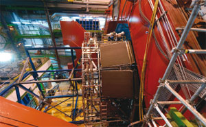
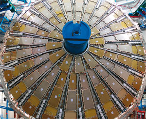
When it starts up the ALICE experiment will observe collisions of heavy ions in CERN’s Large Hadron Collider (LHC), where “fireballs” of extremely hot and dense matter will be fleetingly made. Up to 20,000 tracks will emerge from each fireball, and one of the challenges for ALICE will be to identify different particles among this veritable “haystack” (in p20). Different elements in the armoury of particle identification for ALICE are now arriving in the experiment’s underground cavern, beginning with the High Momentum Particle Identification Detector (HMPID), which was installed inside the solenoid magnet on 23 September. This was soon followed by the first elements of the Time of Flight (TOF) system and the Transition Radiation Detector (TRD).
The HMPID will extend hadron identification in ALICE up to 5 GeV/c, complementing the reach of the other particle-identification systems. It is a ring-imaging Cherenkov detector in a proximity-focusing configuration, which uses liquid C6F14 as the radiator medium, while a 300 nm layer of caesium iodide (CsI) on the cathode of a multiwire proportional chamber converts the Cherenkov photons into electrons. This layer is divided into 161,280 pads, each 8 mm square, which are individually read out by two ASIC chips, GASSIPLEX and DILOGIC, developed with the Microelectronics Group at CERN.
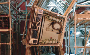
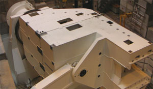
The complete HMPID, realized by Bari University and INFN, CERN (PH-DT1, -DT2 and -AIT groups) and the Institute for Nuclear Research, Moscow, is approximately 8 m wide by 8 m tall, and weighs about 5 t. It comprises seven identical modules shaped to fit against two sides of ALICE’s octagonal magnet. The modules, fully equipped with electronics, were individually transported to ALICE and mounted on a support structure. The complete HMPID was then lowered into the cavern and inserted inside the magnet. Three months of preparation by CERN (PH-DT1 and AIT) and Bari groups, and the help of the CERN transport service, ensured that transport and installation were accomplished within a few hours.
With an active area of about 11 m2 covered with CsI, the HMPID is the largest application of this technology. Development began at CERN in the RD26 project, and it took 15 years for the method to reach the current scale and efficiency. The full production of the 42 photocathodes required to equip the detector, from CsI deposition to quality control, was done by the groups at CERN.
The first week of October saw the installation of the first two supermodules for the TOF system, which will be used to identify the thousands of pions, kaons and protons produced in each fireball. Its basic element is a multigap-resistive-plate-chamber (MRPC) strip, with a 120 cm × 7.4 cm active area made of a sandwich of resistive glass sheets (0.4 mm thick) and spacers, with 96 readout pads, each 3.5 cm × 2.5 cm. The full detector, which contains 1638 MRPC strips with a total of 157,248 readout channels, covers a cylindrical surface of about 150 m2 at 3.7 m from the beamline, and weighs 25 t. It is the responsibility of the INFN sections in Bologna and Salerno, in collaboration with the Institute for Theoretical and Experimental Physics, Moscow, and Kangnung National University, Republic of Korea.
The TRD must identify high-energy electron pairs generated in the fireballs. It comprises 18 supermodules that form a cylinder around the large Time Projection Chamber in the central barrel of the ALICE experiment. Each supermodule is about 7 m long and comprises 30 drift chambers in six layers. The construction of the modules is a collaboration between the Universities of Frankfurt and Heidelberg, GSI Darmstadt, the National Institute of Physics and Nuclear Engineering, Bucharest, and the Joint Institute for Nuclear Research, Dubna, with the radiators produced at the University of Munster.
During the summer the drift chambers for the first supermodule were equipped with readout electronics and inserted into the supermodule hull at the University of Heidelberg. After transportation to CERN on 27 September, the module was tested on the surface using cosmic rays before being lowered into the ALICE cavern on 9 October. The final installation took place a day later.
A new tungsten monocrystalline positron target has generated an intense positron beam at the injector linac of the KEK B-factory (KEKB). It has operated stably since its first use in September 2006 and it is helping to increase the integrated luminosity of KEKB. Crystal positron sources of this kind could be important for the next generation of B-factories and electron–positron linear colliders.
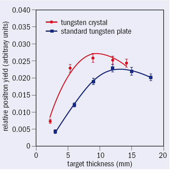
The new positron target at KEKB consists of a 5 mm square tungsten monocrystal 10.5 mm thick, which is bombarded with 4 GeV electrons. The positrons created are then collected and accelerated in succeeding sections up to the final energy of 3.5 GeV for injection into the KEKB positron ring. A conventional target of a 14 mm thick tungsten plate has previously been used, giving a conversion efficiency – the ratio of the number of positrons captured in the positron-capture section and the number of the incident electrons (Ne+⁄Ne–) – of 0.20 (mean). Replacing the tungsten plate with the tungsten crystal has increased the conversion efficiency to 0.25 (mean) (figure 1), which in turn has boosted the positron intensity to its highest since KEKB began operating in 1999.
In a positron source, electrons radiate photons when they interact in a suitable target, and the photons then create electron–positron pairs. The use of a crystal target as a good alternative positron source was first proposed by Robert Chehab and colleagues at Laboratoire de l’Accélérateur Linéaire (LAL), Orsay, in 1989. The method has the advantage of producing high photon intensities by channelling radiation and coherent bremsstrahlung. Experiments at CERN (WA103) and KEK confirmed that the positron yield from a crystal target is remarkably enhanced at higher electron energies. Studies have since been done at KEK to find the optimum crystal thickness as a function of the incident electron energy (figure 2), and Tomsk Polytechnic University has developed tungsten crystals of various thicknesses.
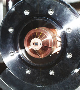
Technology for mounting the tungsten crystal in the positron production station at KEK has also been studied carefully because the <111> crystal axis must be oriented with respect to the direction of the incident electrons to within 1 mrad. To achieve this precise orientation without alignment devices, the target assembly (figure 3) was carefully fabricated, using X-ray Bragg-reflection measurements to ensure that the crystal axis orientation was correct. The team then installed the crystal target at the operational positron source of the KEKB injector linac, and it has since been operating stably. Continued operation at KEKB will provide useful information about radiation damage and the stability of the crystal target.
• This work has been done through the collaborative efforts of Tokyo Metropolitan University, Kyushu Synchrotron Light Research Center, Tomsk Polytechnic University, LAL and KEK.
In 1945 William Hansen at Stanford University built a disk-loaded linear accelerator that produced 4.5 MeV electrons and was less than a metre long. This first electron linac ran at the previously unimaginable frequency of 3 GHz and was so short because it used pulsed high-power klystrons invented by the Varian brothers and developed during the Second World War. Hansen had aimed to advance research in nuclear physics, but his invention was to have an enormous impact on medicine. By the 1970s the company Varian led the market in producing what is now “conventional” radiotherapy systems based on the same type of linac running at the same frequency.
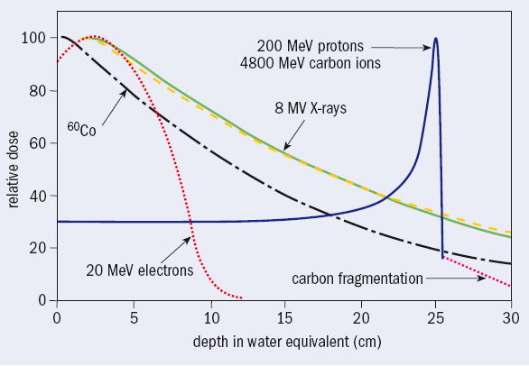
In developed countries every year some 40,000 per 10 million inhabitants are diagnosed as having cancer, around half of whom are treated with high-energy photons produced by electron linacs. There are almost 10,000 electron linacs worldwide, which run more than one shift a day. They irradiate around 4 million patients a year, each in about 30 sessions over 5–6 weeks. The photon beams have energies of a few million electron volts, but are still called X-rays by medical doctors. They have replaced low-energy X-rays and the gamma radiation from radioactive cobalt because they deposit the dose (the energy per unit mass) at greater depth (see figure 1).
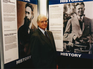
In the same year of Hansen’s invention, and not far away, Robert R Wilson – a Harvard associate professor who was working on cyclotrons with his old teacher Ernest Lawrence at the Radiation Laboratory in Berkeley – was computing the shielding thickness for a 150 MeV cyclotron to be constructed and installed at Harvard. Fifty years later, opening the Advances in Hadron Therapy conference, held at CERN in 1996, Wilson said, “I found that a few inches of lead would fix everything. But I did not stop. Why? Fifty years later I do not know why I did not stop. I suppose the first reason was just plain simple curiosity. So I went on and I jumped into the most obvious thing I could do next: because one could hurt people with protons, one could probably help them too. So I tried to work out every detail and I was surprised to see that the Bragg curve came up and came down very sharply,” (Wilson 1997). The narrow Bragg peak at the end of the range (figure 1) prompted him to publish in the journal Radiology a now-famous paper suggesting the use of protons (and carbon ions) to irradiate tumours while sparing – much better than with X-rays – the healthy tissue traversed, contiguous and located more deeply (Wilson 1946).
However, the resonance within the medical community was almost zero and it was a decade before Berkeley and Harvard treated patients with proton beams from accelerators originally designed for nuclear-physics experiments. It wasn’t until the beginning of the 1990s that radiation oncologists started to recognize this new therapeutic method, because the apparatus was huge by medical standards and the irradiations were done in nuclear-physics laboratories with horizontal particle beams and simple beam-shaping methods. By 1993 about 10 000 patients worldwide had been treated with protons, and by the end of 2006 this has reached 50,000. Today five companies supply turnkey proton-therapy centres.
It is no surprise that from 1961 interesting clinical results for proton therapy were obtained at Harvard where radiotherapists at Massachusetts General Hospital and physicists from Harvard have successfully treated many thousands of head and neck tumours. Eventually in 1993 at the Loma Linda University Medical Center in California, the first proton synchrotron dedicated to proton therapy began irradiating patients in three treatment rooms featuring magnet beamlines on 10 m high gantries, which rotate around the patient. Again, it is no surprise that the Loma Linda synchrotron was built at Fermilab, the particle-physics laboratory that Wilson created and then directed until 1987.
Carbon ions join the fight
Heavier ions than protons, such as helium and later argon, first came into use at Berkeley in 1957 and 1975, respectively. At the old 184 inch cyclotron 2800 patients received brain treatment with helium beams: the lateral spread and range straggling are smaller and this leads to much better dose gradients than protons. At the Bevalac, argon beams were tried to increase the effectiveness against hypoxic and otherwise radio-resistant tumours, i.e. tumours that need deposited doses 2–3 times higher if they are to be controlled with either photons or protons. But problems arose owing to non-tolerable side effects in the normal tissues. After a few irradiations, the Bevalac used lighter ions, first silicon ions and then neon, for 433 patients before it shut down in 1993.
The transition from protons to heavier ions adds another order of magnitude to the complexity of patient irradiation. In the beginning at Berkeley the increase in the relative biological effectiveness (RBE) for ions with respect to photons was believed to be related to the physical parameters of the beam, being the same for different tissues. Since 1980 a large programme of systematic studies of RBE has been carried out at various accelerators, such as Unilac (Darmstadt), Ganil (Caen), Bevalac (Berkeley), the Tandem Van de Graff (Heidelberg) and, later, SIS (Darmstadt). This research studied the effects on very different biological objects, from sub-cellular systems, such as DNA and chromosomes, to biological systems that are resistant to extreme environmental conditions and are used in space research.
The experiments used more than 100,000 biological samples and ion beams from very light to very heavy elements. The research identified the systematic dependence of RBE on physical and biological parameters – mainly the capacity of cells to repair DNA damage – as the most important factor, which was then theoretically modelled for use in treatment planning. In particular, the work showed that for beams of carbon ions the section of the particle track with increased RBE coincides with the few centimetres up to the Bragg peak, while for lighter ions it is concentrated in the last few millimetres. For heavier ions, such as the argon, silicon, and neon ions used previously at Berkeley, it causes significant damage in the normal tissues before the tumour.
For these reasons, in 1994 the synchrotron facility led by Yasou Hirao at the Heavy Ion Medical Accelerator in Chiba (HIMAC), of the National Institute for Radiation Sciences in Japan, treated the first patient with carbon ions, although the accelerator complex was originally designed for ions up to argon.
While an energy of 200 MeV is needed to reach deep-seated tumours (about 25 cm of water equivalent) for protons, 4800 MeV is needed for carbon ions, 24 times higher. Protons beams are obtained either from cyclotrons (normal or superconducting) or from synchrotrons with a diameter of 6–8 m. Currently only synchrotrons are used to produce carbon ions up to 400–430 MeV/u. Their magnetic rigidity is about three times larger than for 200 MeV protons, so synchrotrons of 18–25 m diameter are needed.
Since the end of the 1990s, newly built proton-therapy centres feature isocentric gantries to improve treatment conformity. These avoid high doses to healthy tissue by rotating the beam around the patient as in X-ray treatments. These complex hi-tech systems could not be designed and run effectively and continuously – as is necessary in a hospital environment – were it not for decades of colliding particles and understanding the subatomic world.
Until 1997 relatively simple passive spreading systems were used to produce a spread-out Bragg peak in all hadron-therapy centres. A first scatterer widens the pencil beam while the energy is adapted to the further side of the tumour by appropriate absorbers. More recently, GSI and PSI have developed novel active spreading systems (Haberer et al. 1993 and Pedroni et al. 1995, respectively), which magnetically guide the charged hadrons over the treatment area and modulate the intensity (Intensity Modulated Particle Therapy, or IMPT). All future centres will feature such systems. In particular the ion-therapy centres currently being built at Heidelberg and Pavia have been equipped with the first active beam-delivery system for carbon ions, which restricts the physically and biologically effective end of the track to the target volume (Rossi 2006).
Treating patients
By the beginning of 2006, around 45,000 patients had been treated with proton beams in 12 subatomic physics laboratories and in more than 10 hospital-based proton-therapy centres. (The Particle Therapy Co-ordination Group updates the number of patients treated at see http://ptcog.web.psi.ch Another 10 centres are running or are being built. This shows that proton therapy is booming. At the same time around 2200 patients have been treated with carbon ions at HIMAC, and about 300 at the pilot project proposed by Gerhard Kraft and built at GSI in Darmstadt.
In a conventional treatment with photons of a few million electron volts, a total dose of 60–70 Gy (1 Gy = 1 J/kg) is deposited in a tumour in typically 30 fractions over six weeks. This “fractionation” gives time for re-oxygenation of hypoxic – and therefore radio resistant – tumour cells and allows them to change from radio-resistant stages in the cell cycle to more sensitive stages. In addition, unavoidably irradiated healthy cells have a chance to repair themselves. A proton treatment typically needs the same number of fractions, but allows higher doses to the tumour and thus larger control rates. A larger dose is beneficial because even a 10% increase in the deposited dose generally increases the probability of local control of the tumour by 15–20%.
With carbon ions, the clinical results from Japan and Germany on head, lung, liver and prostate tumours confirm the radiobiological predictions that they have a larger RBE than protons, because their ionization is 24 times higher, which produces multiple double-strand breaks of the DNA of the traversed cell. This damage cannot be repaired, so ion beams are most suited for slow-growing tumours, which are precisely those tumours that are resistant to photons and protons. It is important to note that, since there is only little repair to damage by carbon ions, the fractionation of the dose is not needed as far as tumour inactivation is concerned, but for the normal tissue in the entrance channel fractionation helps to repair the less severe damage. In principle a patient can be treated in 5–10 sessions, reducing both psychological and financial cost. A proton treatment costs 2–3 times more than a conventional treatment, averaging in the West around €6000, but the economy of carbon treatment is different because the shortening of the treatment allows for effective use of the infrastructures. If confirmed by the ongoing clinical trials, this will reduce the cost of treatment and may become one of the main reasons behind any rapid expansion of light-ion therapy in the future. In addition, having little or no side effects reinforces the necessity of active beam-delivery systems for carbon ions, to tailor the dose to the tumour.
In summary, research indicates that carbon-ion beams should be used in the treatment of deep-seated tumours, which are radio resistant both to high-energy photons and to protons. These tumours are thus the targets of choice in a carbon-ion facility, while proton therapy is well adapted to the cases in which a tumour is close to critical organs that cannot be irradiated (Amaldi and Kraft 2005). Groups of radiotherapists in Austria, France, Germany and Italy have applied specific criteria for each tumour site to the national data and made detailed analyses of the number of potential patients (Carbon-ion therapy 2004). The results of these different approaches are consistent. They show that about 1% of the patients treated today with X-rays should be irradiated with protons as the outcomes are definitely better than conventional therapy. In addition, about 12% of X-ray patients would benefit from proton treatment but further clinical trials are needed to quantify the clinical advantages site by site. Lastly, about 3% of X-ray patients would benefit from carbon-ion therapy, but more clinical trials and dose-escalation studies are needed.
Overall, 15% of the approximately 20,000 patients per 10 million inhabitants treated with conventional radiation would receive better treatment with hadron beams. Irradiating these patients would require 3–4 proton treatment rooms (i.e. a centre treating about 1500 patients a year) per 5 million people and a carbon-ion centre per 35 million people. A balanced national programme can therefore make good use of dual centres that accelerate carbon ions and protons and feature fixed ion beams (horizontal, vertical and inclined) and rotating gantries for protons.
Hadron therapy in Europe
In the past five years Europe has made important steps in developing and building hospital-based dual centres for carbon ions and protons. Based on the success of GSI’s pilot project, the Heidelberg Ion Therapy Centre (HIT), designed by GSI, was approved in 2001 and civil engineering work began in November 2003. This centre features two horizontal beams and the first carbon-ion rotating gantry, which is 25 m long and weighs 600 tonnes. The first treatment will be at the end of 2007.
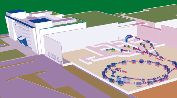
At the end of 1995 Ugo Amaldi, with Meinhard Regler of the Med-Austron project, attracted CERN management’s attention to the design of an optimized synchrotron for light-ion therapy. This was the starting point of a five-year Proton and Ion Medical Machine Study (PIMMS) (Badano et al. 1999 and 2000). As a development of this initiative, in 2002 the Italian health minister financed a second European centre, based on the PIMMS design modified by the TERA Foundation (Fondazione per Adroterapia Oncologica). This is now being built in Pave by the Centro Nazionale di Adroterapia Oncologica Foundation (CNAO) with strong support from INFN (figure 3). It will be ready by the end of 2007.
At the end of 2004 the Austrian authorities approved the Med-Austron project, granting a substantial part of the required funding for the construction of a dual centre in Wiener Neustadt. The tendering procedure to acquire a turnkey carbon-ion facility is now almost complete. Similarly, in May 2005 the French government approved the ETOILE project to be built in Lyon.
In 2002 the initiatives at Heidelberg, Lyon, Pave, Stockholm (where the Karolinska Institute has proposed a similar facility) and Wiener Neustadt all teamed up with the European Society for Radiotherapy, CERN and GSI to form the European Network for Light Ion Therapy, which the European Union financed for three years. The work by this network, and the existence of its potential successor, guarantees that carbon-ion therapy in Europe is on the right track and that the foreseen facilities will be run for the benefit of all European patients. During 2006 a larger group of institutes and hospitals from 15 countries has come together to prepare a new proposal for the EU Framework Programme FP7 under the name ENLIGHT++ (CERN Courier June 2006 p27).
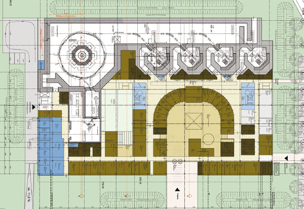
In addition, in January 2006 contracts for a privately financed carbon/proton centre were signed by Rhön–Klinikum–AG, which owns more than 40 German hospitals, including the Giessen-Marburg University clinics, and Siemens Particle Therapy. When it starts up in 2010, the new heavy-ion therapy facility in Marburg (figure 4) will show that hadron therapy with ion and proton beams has left research and arrived in the clinical environment.
Future developments
The unique physical and biological properties of hadron beams are better for patients than the most recent photon image guided radiotherapy (IGRT) techniques if the position of the tumour target can be accurately determined and an active irradiation system can follow the movements of the treated organ. To achieve this, two approaches have been considered. One uses feedback systems that redirect the moving beam during scanning, while the other uses the multiple “repainting” of the target to avoid the local delivery of larger or smaller doses than predicted. In the former, the online motion correction can be done in 3D, using the scanning system for the lateral correction and a fast passive absorber for the depth correction. Experiments at GSI with a phantom showed that the homogeneity and the steep gradients can be preserved to 95% compared with static target irradiation. PSI will pursue the latter approach with proton beams at the PROSCAN project.
Scientists at KEK and TERA have proposed two types of fast cycling accelerators, better suited than cyclotrons and synchrotrons to treating moving organs. They are, respectively, the fixed field alternating gradient accelerator (a mixture of a cyclotron and a synchrotron) and the cyclinac (the combination of a low-energy high-current cyclotron and a high-frequency linac), both of them suitable for proton and carbon-ion therapies. It will take a few years to see what the best solution is both economically and technically.
Without waiting for these developments, industry has shown interest in the upcoming market of hadron therapy, proposing solutions based on synchrotrons and cyclotrons. Five companies already sell proton-therapy units. In the heavy-ion market Mitsubishi has designed a micro-HIMAC, a synchrotron for combined proton and carbon therapy, while Siemens Particle Therapy offers a combined proton/carbon facility on the basis of exclusive licences of the GSI patents and know-how. At present a commercial company is discussing the licensing of the PIMMS/CNAO synchrotron design with the CNAO Foundation. Moreover scientists at the INFN Laboratori Nazionali del Sud in Catania have designed a 300 MeV/u superconducting cyclotron that accelerates hydrogen molecules and carbon ions, allowing treatment with protons of all tumours as well as treatment with carbon ions of tumours located at a water depth of about 15 cm. The Ion Beam Application company in Belgium has transformed this design into a commercial product. The recent interest of industrial companies in ion therapy indicates its large potential, which has its roots in the instruments developed for fundamental research in subatomic physics.
In Europe, one in three people is expected to confront some form of cancer during his or her lifetime. In Switzerland alone, this amounts to about 28,000 patients a year – 70% of whom undergo radiotherapy, now the second most successful form of treatment after surgery. While the majority of tumours are treated with photons, the use of proton beams, first proposed 60 years ago, is becoming increasingly important for deep-seated tumours, as the technique moves from accelerator centres to dedicated clinical facilities. The Paul Scherrer Institute (PSI) is one of Europe’s leading centres for proton-therapy research and has recently begun work with a new superconducting proton cyclotron, COMET, and beamline to serve its proton-therapy project, PROSCAN.
PROSCAN grew from PSI’s decision in 2000 to expand its radiotherapy activities by developing a new compact gantry system to enable the laboratory’s successful spot-scanning technology to be used in a hospital environment. The project also aims at optimizing treatment methods and expanding the spectrum of treatable forms of cancers, as well as transferring the technology and know-how to industry and other radiotherapy centres, including the education and training of specialist personnel.
PSI’s pioneering development of the spot-scanning technique for deep-seated tumours dates back to its former times as the Swiss Institute for Nuclear Research (SIN), when the laboratory established a therapy programme using pions. This used a dynamic beam-delivery system of 60 converging pion beams where the patient’s target area could be moved 3-dimensionally within a body (bolus) of water. Some 500 patients were treated with this system between 1981 and 1992. Protons for both the pion work and the subsequent proton-therapy programme at the Gantry 1 system described below were produced by the main 590 MeV Ring Cyclotron and a beam-splitter system, which at the same time served the main users in particle physics and materials science. In 1984, in collaboration with the Lausanne University Eye Clinic, the very successful OPTIS Proton Therapy Programme began. A first in Europe, this facility has treated more than 4400 patients with differing forms of eye tumours, using a 70 MeV horizontal proton beam from PSI’s Injector 1 cyclotron. With a success rate of better than 98%, it continues to treat the highest number of eye patients worldwide each year, and has led in turn to the establishment and operation of six new European facilities in England, France, Germany, Italy and Sweden.
Modern ways to achieve a conformal dose distribution tailored 3-dimensionally to a target tumour volume, while sparing healthy tissue, use active scanning rather than conventional passive scattering (Goitein et al. 2002). The latter, which has fewer degrees of freedom, uses a set of scattering foils to produce a laterally spread-out proton beam of constant range. A set of individually manufactured collimators and compensators then contours the beam to match each target volume. The range of the protons is subsequently modulated by, for example, a rotating range-shifter wheel, which alters the proton depth profile.
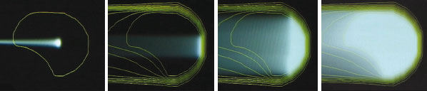
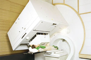
Figure 1 illustrates the principle of active scanning. Here the dose delivery to the patient is achieved through the sequential superposition of single pencil beams of protons, each of which produces a hot spot at the Bragg peak, where the protons deposit most of their energy. The hot spot is about 1 cm3 for a Gaussian beam profile of 7–8 mm FWHM. Lateral scanning is possible either using sweeper magnets or by moving the patient table, or by a combination of both. Depth modulation, on the other hand, is achieved either by a fast active degrader or by changing the beam energy. Combining these options with both a beam-delivery system that can rotate and an eccentrically mounted counter-rotating patient table yields the very compact (4 m diameter) PSI Gantry 1 system (figure 2).
This system allows a dose application of almost 10,000 spots/litre to be applied in a few minutes with an individual spot-dose precision of 1%. It is the only facility in the world to use a dynamic beam-delivery technique based on active spot scanning with protons. A similar scanning system using a horizontal beamline has been developed for carbon ions at Germany’s national laboratory for heavy-ion physics, GSI in Darmstadt.
Gantry 1 was designed to treat deep-seated tumours and since it started up in 1996 has handled around 260 patients, some 69 of whom were treated with a new therapy plan using intensity-modulated proton therapy (IMPT), which further reduces the dose to healthy tissue. One of the main disadvantages of the present spot-scanning technique as applied in Gantry 1 is the modulation speed, which is currently too slow to apply IMPT to moving target volumes. This will soon be addressed with the new features being incorporated into Gantry 2, within the PROSCAN project.
The PROSCAN project
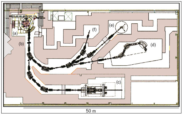
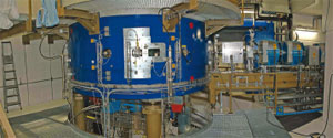
When PROSCAN is complete it will consist of six main features (figure 3), with the newly commissioned 250 MeV superconducting cyclotron, COMET, as the heart of the facility (figure 4). ACCEL Instruments, Germany, built this machine in close co-operation with accelerator specialists at PSI, and based it on a design by Henry Blosser of Michigan State University. It will allow year-round therapy operation. The decision in favour of a cyclotron was based on the benefits of the 100% duty cycle and the need to control the beam intensity precisely and dynamically prior to acceleration. The restriction of the fixed energy from a cyclotron in turn requires a degrader system that acts rapidly, allowing fast, small energy steps to be implemented. This, together with a beam-diagnostics system after the degrader, means that the rapid modulation of both energy and intensity can be fully exploited, allowing the possibility of fast volumetric rescanning and the study of IMPT for moving tumours. The new machine also achieved the stringent requirement of an 80% extraction efficiency and an availability of 98%.
Gantry 1 will remain unchanged and will continue as the workhorse for treating patients in PROSCAN. The development work will concentrate on implementing IMPT methods into clinical practice, including treatment planning, dosimetry and quality-assurance. The present OPTIS eye-treatment facility at the Injector 1 cyclotron will continue its successful operation until mid-2007 when it will be transferred to the new 70 MeV area at PROSCAN. This will involve a complete re-design of the control system and treatment procedures. The second of the two horizontal beamlines will do biological and dosimetry experiments.
Finally, to meet the challenge to proton therapy of producing a beam-scanning method that can overcome the sensitivity to organ motion, a new compact gantry system, Gantry 2, is being built to be implemented in 2007, with the first treatments of patients expected in 2008 (figure 5). This will allow faster beam scanning by 2D magnetic deflection, to achieve multiple target rescannings of the same volume within a single sitting (Pedroni et al. 2004).
Once the PROSCAN facility is fully operational it is expected that the number of patient treatments for deep-seated tumours will increase by a factor of 3–4 (150–250 patients a year) with a further 200–300 patients a year benefiting from the OPTIS eye- treatment station. It has taken some 50 years from the basic idea for protons to come of age as a clinical tool, enabling more than 40,000 patients so far to benefit from this therapy developed in a multi-disciplinary fashion. Although the future is clearly aimed at providing dedicated commercial facilities for hospitals and clinics, the present role played by accelerator laboratories such as PSI, in developing new methods and the technology to implement them, is an essential ingredient in achieving this goal.
Researchers from the Technion – Israel Institute of Technology have used the Accelerator Test Facility (ATF) at the US Department of Energy’s Brookhaven National Laboratory to demonstrate the feasibility of particle acceleration by stimulated emission of radiation (PASER). This is in effect a particle analogue of the laser process.
Levi Schächter at Technion initially demonstrated theoretically the concept behind PASER. Its essence relies on the possibility of transferring energy stored in an active medium (with excited atoms) directly to electrons interacting with the medium, and thereby increasing their energy.
In lasers, photons traversing an active medium stimulate the atoms via collisions so that the atoms give up their excess energy as additional photons forming a coherent beam. In PASER, the atoms in the active medium transfer their energy directly to an electron beam in a coherent way.
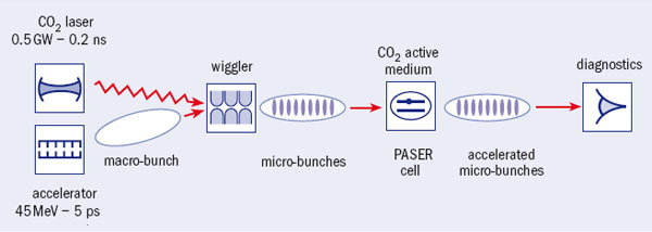
To reach significant acceleration in a PASER, a macro-bunch of electrons injected into the PASER cavity should be modulated, forming a train of micro-bunches with a periodicity identical to the resonance frequency of the medium. In other words, coherent collisions of the second kind between the electrons in the train and the excited molecules should occur.
In the proof-of-principle experiment by Samer Banna, Valery Berezovsky and Schächter, electrons in a macro-bunch with an energy of 45 MeV were modulated by their interaction with a high-power CO2 laser in a wiggler, to make a train consisting of about 150 micro-bunches, each several femtoseconds long. About 15% of the electrons in the train collected at the spectrometer at the end of the PASER cavity had absorbed energy stored in the cavity, increasing the total kinetic energy of the macro-bunch by about 0.15%.
Accelerating electrons in this way provides new opportunities as the effective quality factor of such a cavity may become comparable to that of macroscopic superconducting cavities. In particular it will be a challenge to try to use this technique to generate ultra-low-emittance beams.
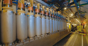
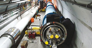
The first cryogenic feedbox designed to supply electricity to the superconducting magnets for one of eight arcs has been installed at Point 8 of the Large Hadron Collider (LHC). This milestone is the precursor to the cool-down of sector 7-8, scheduled for the coming months. Researchers will position a total of 16 such feedboxes at either end of the eight arcs, forming the ends of the continuous sections of cryostat. Each one weighs 12.7 t, is 10 m long and must withstand a pressure of 0.25 MPa.
Power leads, the lower extremities of which are immersed in liquid helium, bring the electrical power from room temperature to cryogenic temperature. Helium gas actively cools them and is injected at their base at 20 K and comes out at room temperature at the top. The power leads use ceramic high-temperature superconductor to limit the heat loads – the first time that these materials have been used on this scale.
The power supply to the LHC’s straight sections requires smaller electrical feedboxes. There will be 44 feedboxes around the LHC ring, equipped with 1200 current leads carrying 120–13,000 A.
Meanwhile, on 5 September the 1000th cryomagnet (superconducting magnet system) was installed between Point 3 and Point 4. During the same week, the final cryomagnet for sector 8-1 was also installed. There are 1746 cryomagnets, of which 1232 are the famous dipoles.
While most of the LHC experiments are on a grand scale, LHC forward (LHCf) is quite different. Unlike the massive detectors that are used by ATLAS or CMS, LHCf’s largest detector is a mere 30 cm. Rather like the TOTEM detector, this experiment focuses on forward physics at the LHC. The aim of LHCf is to compare data from the LHC with various shower models that are widely used to estimate the primary energy of ultra-high-energy cosmic rays.
The LHCf detectors will be placed on either side of the LHC, 140 m from the ATLAS interaction point. This location will allow the observation of particles at nearly zero degrees to the proton beam direction. The detectors comprise two towers of sampling calorimeters designed by Katsuaki Kasahara from the Shibaura Institute of Technology. Each is made of tungsten plates and plastic scintillators of 3 mm thickness for sampling.
Yasushi Muraki from Nagoya University leads the LHCf collaboration, with 22 members from 10 institutions and four countries. For many of the collaborators this is a reunion, as they had worked on the former Super Proton Synchrotron experiment UA7.
September saw the completion in the underground cavern of the first of the big wheels for the ATLAS muon spectrometer. The muon spectrometer includes four large wheels at each end of the barrel part of the detector, each measuring 25 m across. Six of these wheels will be composed of thin-gap chambers for the muon trigger system – the first wheel is one of these – while the other two will be made of monitored drift tubes to measure the position of the muons.
POSIPOL 2006, an international workshop that was held earlier this year at CERN, was dedicated to the production of polarized positrons using the Compton back-scattering of a high-power laser beam by electrons of a few giga-electron-volts. The particular focus was on applications to the two future linear-collider studies, the International Linear Collider (ILC) and the Compact Linear Collider (CLIC). The workshop, which attracted around 50 experts from Europe, Asia and the Americas, was jointly organized by the CLIC team at CERN, the European CARE-ELAN network, the Japanese high-energy accelerator research organization, KEK, and the Laboratoire de l’Accélérateur Linéaire (LAL) at Orsay. It led to a roadmap and a series of recommendations for future R&D on positron sources for linear colliders.
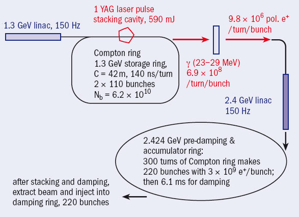
Polarized positrons (POSItons POLarisés in French, hence POSIPOL) are produced by bombarding a tungsten target with polarized photons. The latter are generated either from a helical undulator or from the scattering of a polarized high-power laser beam with an unpolarized high-energy electron beam. For this second scheme, requirements on the intensity of both the electron beam and the laser beam are significantly relaxed by stacking the laser beam in an optical cavity with an enhancement factor of up to 1000, and by re-using the electrons, which are stored in a so-called Compton ring. This scheme also implies the stacking of the produced positrons in a storage ring. Control of the laser system and of the high-quality optical cavity is crucial, as is the electron-beam dynamics in the presence of electron-laser collisions. The various aspects of this scheme, as well as comparisons with the undulator method, formed the main topics for the sessions at the workshop.
Robert Aymar, CERN’s director-general, opened the workshop, and was followed by Louis Rinolfi, POSIPOL chair, who set the scene with a look at the state-of-the-art for producing polarized positrons and a reminder of the scope of the workshop. In the first overview session, Gudrid Moortgat-Pick from CERN stressed the importance of positron polarization for future linear colliders. Both a Compton source and an undulator scheme are being considered for the ILC, as described by KEK’s Junji Urakawa and John Sheppard of SLAC respectively. Frank Zimmermann of CERN presented a proposal for a Compton source for CLIC, demonstrating that the pertinent requirements are much less demanding than they are for the ILC. He also emphasized the large synergy with ongoing developments for a Compton ring for medical applications.
Several talks by Susanna Guidicci of Frascati, Alessandro Variola of LAL, and Eugene Bulyak and Peter Gladkikh of the Kharkov Institute for Physics and Technology (KIPT) discussed the beam dynamics and optics designs for Compton rings. One suggestion was to use a pulsed mode of operation for the Compton ring with a specific technique for radio frequency (RF) phase modulation. Vitaly Yakimenko of Brookhaven National Laboratory described the merits of an alternative single-pass Compton scattering approach involving a high-duty-cycle electron linac and a battery of CO2 lasers.
Several talks addressed advances in laser systems, including those by Igor Pogorelsky of Brookhaven, Brent Stuart of Lawrence Livermore National Laboratory and Sudhir Dixit of Oxford. Yoann Zaouter of Amplitude Systems highlighted the dramatic evolution of fibre lasers over the past decade. Products from Time-Bandwidth, presented by Thomas Ruchti, achieve parameters close to what is needed.
Tsunehiko Omori of KEK presented the first experimental results on polarized positron production using Compton back-scattering at KEK’s Accelerator Test Facility (ATF), which have recently been published in Physical Review Letters. The E-166 undulator experiment at SLAC also has results, as described by Andreas Schaelicke of DESY/Zeuthen.
Ian Bailey of the UK’s Cockcroft Institute discussed aspects of positron production, in particular targets, and described the development of a conversion target. Vladimir Strakhovenko of the Budker Institute of Nuclear Physics (BINP) presented theoretical calculations of the radiation spectrum and photo-production in a target. Robert Chehab of IN2P3/Lyon and Wei Gai of Argonne National Laboratory addressed the positron production process, in particular matching and capturing positrons downstream of the target. Masao Kuriki of KEK talked about systems considerations for the positron source at the ILC, in particular construction, commissioning and availability of undulator and Compton schemes.
In the session on equipment and diagnostics, Fabian Zomer of LAL discussed ongoing studies of non-planar optical resonators, and Peter Schueler of DESY looked at beam polarimetry issues. A final R&D session focused on optical cavities. Viktor Soskov of the Institute for High Energy Physics, Moscow, reviewed R&D on a high-finesse cavity at LAL, and Hiroki Sato of Hiroshima described R&D on an optical cavity for the ILC. Kazuyuki Sakaue of Waseda University, Tokyo, described the experimental plan for X-ray generation using a pulse-stacking optical cavity.
At the end of the workshop, POSIPOL participants identified a number of critical issues that still need to be demonstrated, both for positron sources that are based on an undulator scheme and for the alternative Compton scattering scheme. The two approaches are in principle equivalent, but there are differences in the photon spectrum, photon energies, angular photon spectrum, power on the conversion target, collimation efficiency, operational efficiency and implementation cost. For the Compton scheme, the photon and positron yields must be simulated with a realistic lattice and energy spread. Many items require further study, including laser systems, 6D positron distribution, stacking in a pre-damping ring, required RF power, Touschek lifetime, beam instabilities and the heat-load limit of optical cavities.
The workshop also agreed on a roadmap for future R&D to address the common issues in Compton and undulator sources. Noteworthy common recommendations for the two schemes concern the analysis of the systematic errors in the polarization measurements, the comparison of yields and polarization, the optimization of pre- and post-selection of positrons and the evaluation of the cost.
For the undulator scheme, the main recommendations were the publication of E-166 results, the evaluation of emittance degradation in the undulator and the technical demonstration with an undulator several metres long. For the Compton scheme, the main recommendations were the publication of the design of a Compton ring with a chicane and with optimization of the energy of the Compton photons, the development of a reliable power laser taking into account the polarization, the simulation of stacking into a damping ring, the comparison of a single-pass scheme with the ring scheme and the comparison of CO2, YAG and fibre lasers.
The workshop also addressed validating design choices and demonstrating feasibility in experiments at KEK’s ATF, Brookhaven’s ATF and the DAFNE storage ring at Frascati. In May, the KEK–ATF Technical Board approved an experimental programme of installing and operating laser pulse-stacking cavities in the ATF damping ring during 2006 and 2007. The goal is the simultaneous demonstration of the high enhancement factor that is required by POSIPOL, the small laser spot and a small beam–laser collision angle in multi-bunch operation. Optimized optical cavities from LAL may be installed later at the KEK-ATF, enabling the study of high-intensity multi-bunch gamma-ray generation by Compton scattering.
Another new project at KEK would allow accumulation experiments with electron beams. An experimental optimization for the Compton source inside a laser cavity is also foreseen at the Brookhaven ATF, and single-pass Compton collisions could be tested with the drive beam of the CLIC Test Facility 3 at CERN.
Since the POSIPOL workshop, LAL has written a letter of intent concerning R&D activities on polarized positron sources. The idea is to submit a proposal as a Joint Research Activity (JRA–POSIPOL) in the context of the European Framework Programme 7. Several institutes have already expressed their interest: LAL, INFN/Frascati, CERN, DESY Zeuthen, the Institut de Physique Nucléaire de Lyon, BINP, the National Science Center KIPT, Université-Paris-XI, KEK, Waseda University and Kyoto University.
For the first time, on 18 August, the OPERA detector in the Gran Sasso Laboratory in Italy recorded the interactions of neutrinos sent from CERN, 732 km away. During the following 11 days, OPERA had about 120 hours of beam time and identified around 300 interactions correlated with the neutrino beam, in-line with predictions. This marked both a successful culmination for the beam-commissioning phase of the CERN Neutrinos to Gran Sasso (CNGS) project and a promising start to data-taking for OPERA. The ultimate aim is to observe oscillations from muon neutrinos into tau neutrinos.
The CNGS beam line incorporates a target to produce pions and kaons and a magnetic horn and reflector to focus them before they decay to muons and neutrinos in a 1 km vacuum pipe. At the end of the decay pipe, a barrier of graphite and iron absorbs the remaining hadrons, leaving only muons, which are quickly absorbed downstream in the rock, and a beam of neutrinos travelling towards Gran Sasso.
The CNGS team began gradually to commission the installation at the start of 2006, beginning with tests without beam of some 200 items of equipment. Then, from 10 July, protons from CERN’s Super Proton Synchrotron were sent step-by-step through the beam line to the target, using a beam 100 times less intense than the nominal beam. After analysis at the end of July, commissioning resumed at high intensity.
The OPERA Collaboration, comprising 170 physicists from 35 research institutes and universities worldwide, began installing their detector in the underground laboratory at Gran Sasso in 2003. The detector has two identical Super Modules, each containing a target section and a large-aperture spectrometer. The target consists of alternate walls of lead/emulsion bricks and modules of scintillator strips for the target tracker. The spectrometer, which detects muons emerging from neutrino interactions, consists of a precision drift-tube tracker, a 1.55 T magnet and resistive plate chambers (RPCs) inserted into the magnet.
During this first run, researchers verified the operation of the electronic detectors (4000 m2 of RPCs, 6000 m2 of scintillator strips and two magnets), checked the synchronization of the OPERA and CNGS clocks and tested the algorithms governing the selection of interesting events. CNGS can send at most two bunches of neutrinos every 6 s, and to reduce the background noise, the experiment has to select events exactly as a bunch passes through. The two 10.5 µs bunches are separated by 50 ms, and the design synchronization accuracy between CERN and OPERA is better than 100 ns.
OPERA is now ready to enter the next phase, aimed at observing neutrino interactions in the emulsions of the detector’s 200,000 target bricks, which will be produced and installed over the coming months. The long search for tau neutrinos will start in earnest at the end of October and will last at least five years.
To provide the best experiences, we use technologies like cookies to store and/or access device information. Consenting to these technologies will allow us to process data such as browsing behavior or unique IDs on this site. Not consenting or withdrawing consent, may adversely affect certain features and functions.
Functional
Always active
The technical storage or access is strictly necessary for the legitimate purpose of enabling the use of a specific service explicitly requested by the subscriber or user, or for the sole purpose of carrying out the transmission of a communication over an electronic communications network.
Preferences
The technical storage or access is necessary for the legitimate purpose of storing preferences that are not requested by the subscriber or user.
Statistics
The technical storage or access that is used exclusively for statistical purposes.The technical storage or access that is used exclusively for anonymous statistical purposes. Without a subpoena, voluntary compliance on the part of your Internet Service Provider, or additional records from a third party, information stored or retrieved for this purpose alone cannot usually be used to identify you.
Marketing
The technical storage or access is required to create user profiles to send advertising, or to track the user on a website or across several websites for similar marketing purposes.