A new method for the radiography of coronary blood vessels (known as angiography) promises to make examinations much easier for patients. The NIKOS intravenous angiography technique, which produces an X-ray image of coronary arteries, was developed by the DESY laboratory, Hamburg, in collaboration with doctors from the University Hospital Hamburg-Eppendorf and the Bevensen Heart Centre, and physicists from the University of Siegen. DESY has examined a total of 379 patients from all over Germany and abroad with extremely satisfactory results.
With the successful conclusion of these trials at DESY’s HASYLAB synchrotron radiation centre, a door opens for routine application of the new technique. However, this would have to be at a specially equipped clinic, with a compact source of monochromatic X-rays. An initial design for such a source, based on a storage ring, has already been made at DESY.
The coronary arteries surround the heart and supply it with blood. If they become constricted, a heart attack can result. To look for these life-threatening constrictions (stenoses), doctors normally insert a long catheter into the coronary vessels via the groin and the aorta. They then inject a contrast medium containing iodine through the catheter and make an X-ray. Such invasive examinations can be an unpleasant experience for patients.
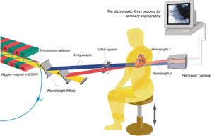
The NIKOS technique eliminates the need for surgical procedures. Instead, the iodine is injected intravenously. Greatly diluted on its journey through the circulatory system, the iodine concentration is so low by the time it reaches the coronary arteries that conventional X-ray tubes cannot produce a clear image. The HASYLAB scientists use intense monochromatic X-rays from the DORIS electron ring as well as a special “two-colour” method to reveal the coronary arteries.

Of the 379 patients examined at DESY using the new technique, 60 underwent a subsequent diagnosis based on conventional X-ray exposure. The diagnoses displayed good agreement.
There are other non-invasive and minimally invasive procedures for imaging the coronary vessels – magnetic resonance imaging (MRI) and electron beam computed tomography (EBCT). However, compared with these methods, the NIKOS technique claims to provide the best image quality. Its output resolution is also better and, unlike MRI, metallic implants do not degrade image quality. However, none of these methods will be capable of replacing conventional coronary angiography in the long term. This is because the conventional method also allows for surgery during the examination, such as repair (angioplasty) or tube implantation (stent).
NIKOS allows the imaging of bypasses and stents in check-ups and postoperative examinations. Further improvements to the system and its associated techniques could increase image quality and therefore the value of the diagnosis.


