Machines made in industry are becoming smaller.
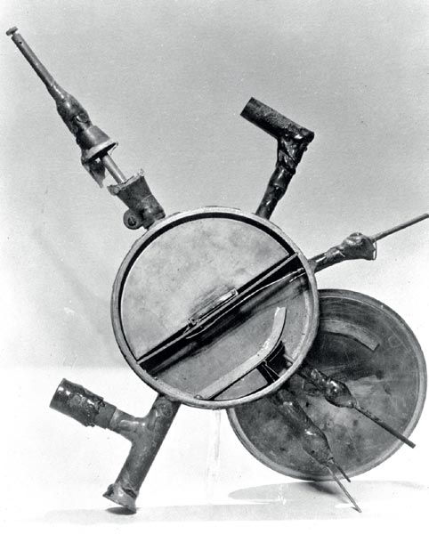
Image credit: LBNL.
The first cyclotron, built in 1930 by Ernest Lawrence and Stanley Livingston, was 4.5″ (11 cm) in diameter and capable of accelerating protons to an energy of 80 keV (figure 1). Lawrence soon went on to construct higher-energy and larger-diameter cyclotrons to provide particle beams for research in nuclear physics. Eighty years ago this month, he and Livingston published a seminal paper in which they described the production of light ions with kinetic energies in excess of 1 MeV using a device with magnetic pole-pieces 28 cm across (Lawrence and Livingston 1932). By 1936 John Lawrence, Ernest’s brother, had made the first recorded biomedical use of a cyclotron when he used the 36″ (91 cm) machine at Berkeley to produce 32P for the treatment of leukaemia. Since then, the physics design of the cyclotron has improved rapidly, with the introduction of alternating-gradient sector focusing, edge focusing, external ion-source injection, electron cyclotron-resonance sources, negative-ion acceleration, separated-sector technology and the use of superconducting magnets.
Historical developments
However, other accelerator designs were evolving even faster, with the construction of the synchrocyclotron, the invention of the synchrotron, of linear accelerators and of particle colliders that were capable of generating the extremely high energies needed by the particle-physics community. The usefulness of the cyclotron appeared to diminish but in 1972 the TRIUMF laboratory in Canada turned on the world’s largest cyclotron, at 2000 tonnes with a beam-orbit diameter of 18 m and negative-ion acceleration. Two years later, in Switzerland, PSI brought into commission a large separated-sector, 590 MeV proton cyclotron. Both of these machines have contributed to isotope-production programmes. More recently, a superconducting ring cyclotron delivering a proton beam energy of 2400 MeV has been built in Japan at the Riken research institute.

Image credit: IBA.
Nevertheless, the value of the cyclotron as a method for producing medical isotopes had come under further pressure in the 1950s and early 1960s from the availability of numerous nuclear-research reactors that had high neutron fluxes, large-volume irradiation positions and considerable flexibility for isotope production. These attributes allowed the generation of important radioisotopes such as 99Mo, 131I, 35S and even 32P more easily and more cost effectively.
Nevertheless, there remained a few radionuclides with neutron-deficient nuclei that were important for medical imaging but could be produced only by particle accelerators. These included 123I and 201Tl, used for nuclear cardiology, and others such as 111In. The production reactions needed were often of the type where a proton knocks out only a few neutrons, (p,xn) with x=1 or 2 or 3, so that the accelerator energy required was usually no more than around 30 MeV. Consequently the use of the medium-energy cyclotron was revived. The first dedicated medical-isotope cyclotron was designed and built at the Hammersmith Hospital in London in 1955 and was followed by dozens of research-based cyclotrons, often with their own bespoke designs.
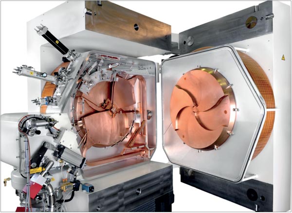
Image credit: General Electric Healthcare.
The routine use of radioisotope-labelled medical products and the demand for radiopharmaceutical injections for patients led to the creation of a new sector of industry: to supply cyclotron systems capable of the production of medical isotopes. Commercial companies started to design, build and supply complete cyclotron systems specifically for this purpose. The first generation of these industrial cyclotrons was made available by companies such as Philips in the Netherlands and The Cyclotron Corporation (TCC) in the US, but these machines were usually complicated instruments requiring considerable physics expertise for operations and maintenance. Second-generation cyclotrons, with more compact designs and improved engineering, were developed later by Scanditronix in Sweden, Thompson CSF in France and Sumitomo and JSW in Japan, all with designs that led to lower radiation doses to the operators. Around 1980, the first negative-ion industrial cyclotron, the CP-42, became available from TCC, with 40 MeV proton extraction.
In 1988, a major step forward occurred with the development by Yves Jongen at the University of Louvain-la-Neuve, Belgium, of an industrial cyclotron customized for medical-isotope production – the Cyclone-30 (figure 2). This new cyclotron was power efficient, had a user-friendly control system and incorporated negative-ion acceleration and charge-exchange stripping for extraction, as developed earlier at the TRIUMF cyclotron. It spawned the start of a new accelerator company in Belgium, IBA SA, and this concept of an optimized industrial design was subsequently adopted by other companies, including Ebco Industries in Canada. Most of these isotope-producing cyclotrons were in the energy range of 20–40 MeV – some having an extracted beam capability of 500 μA or more – and several companies have made available a range of cyclotrons operating at different energies (Schmor 2010).
In addition, a range of positron-emitting, neutron-deficient radio¬nuclides were found to be particularly effective for biomedical human imaging via positron-emission tomography (PET), i.e. 18F, 11C, 15O and 13N. The production energies required for these PET isotopes were lower – from around 5 MeV to 20 MeV – with 18F being the most commonly used. Many of the same industrial companies designed even smaller cyclotrons at around either 17 MeV, for high-output 18F production, or around 11 MeV, for lower, hospital-based 18F production, and some cyclotron designs had radiation self-shielding arrangements. PET had long been an imaging technique used in research but by 1998, the medical regulator in the US – i.e. the Food and Drug Administration – had approved the use of PET imaging for several clinical indications. However, 18F has a half-life of only two hours, which limits delivery to small geographic regions. This led to the building of numerous manufacturing facilities for lower-energy PET cyclotrons. It is estimated that by 2010 the world market for small PET cyclotrons was between 50 and 60 a year.
Although 18F, in the form of 18F-deoxyglucose or FDG, has remained the most commonly used radionuclide for PET, numerous other tracers labelled with 18F are in advanced stages of clinical development and eventual commercialization. However, the process of their drug licensing has been particularly slow. Despite the considerable investment made in R&D and manufacturing of FDG by industry, there is a growing concern that the potential for its use in PET imaging and its implementation in personalized medicine has not been achieved fully. There also exist numerous other tracers that provide good images of the human body, many of which use 11C as the radiolabel. However, 11C has an extremely short half-life of only 20 minutes and it would have to be produced inside the facilities of the smallest general hospitals, as well as at larger research institutions.
The drive towards smaller, hospital-based cyclotrons dedicated to producing small quantities of injectable radioisotopes started back in 1989 when IBA, following on from the success of the Cyclone-30, designed the Cyclone 3D – a 3 MeV deuteron cyclotron for 15O production. Some five models have been delivered, but unfortunately 15O has remained a research tool used primarily for blood-flow studies rather than becoming a regular commercial product with its own pharmaceutical-marketing authorization licence.
Another approach towards reducing the size of the cyclotron was the OSCAR cyclotron, originally designed and delivered in 1990 by Oxford Instruments in the UK, and now distributed by EuroMeV. OSCAR is a 12 MeV, 100 μA superconducting cyclotron with an external ion source. Around eight models with this more complicated design have been delivered.
Latest trends
The concept of producing quantities of PET radionuclides that are suitable for one dose to a single patient was not really addressed until 2009, when Ron Nutt of ABT Molecular Imaging Inc developed a small cyclotron with 7.5 MeV energy, positive ion (i.e. proton) acceleration and an internal target for the production of unit doses of 18F. This cyclotron was designed as a component of an integrated production system that also included targetry, a chemistry system based on microfluidic processing, an online chemistry quality-control system and a methodology for radiopharmaceutical product release. The physics of this cyclotron reverts to the more traditional method of proton acceleration and internal targets to reduce the radiation burden associated with stripping inside negative-ion cyclotrons. Nevertheless, this cyclotron system has established a new strategy of producing unit-patient doses of radionuclides with short half-lives. Moreover, the production can be located in smaller clinical facilities, possibly in remote and rural locations around the world.
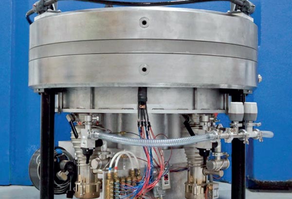
Image credit: ABT Molecular Imaging Inc.
The use of negative-ion acceleration, the ease of charged-particle stripping-extraction and the convenience of having external targets have been preferred by other developers. General Electric Healthcare has recently reported success in the development of a small, vertical cyclotron with a proton energy of around 8 MeV. In Spain, a public–private consortium has announced a development project called AMIT, which is funded by the Spanish Centre for Industrial Technology Development. Within this consortium, the accelerator institute CIEMAT in Madrid will be delivering a cyclotron with a low-energy proton beam. A collaboration has been set up between CIEMAT and CERN to design and build the smallest-possible cyclotron using superconducting technology with a proton energy of around 8 MeV, the objective being to produce single-patient doses of both 18F and 11C in particular. This collaboration with CERN will include the use of some of the accelerator technology and expertise used in building the LHC.
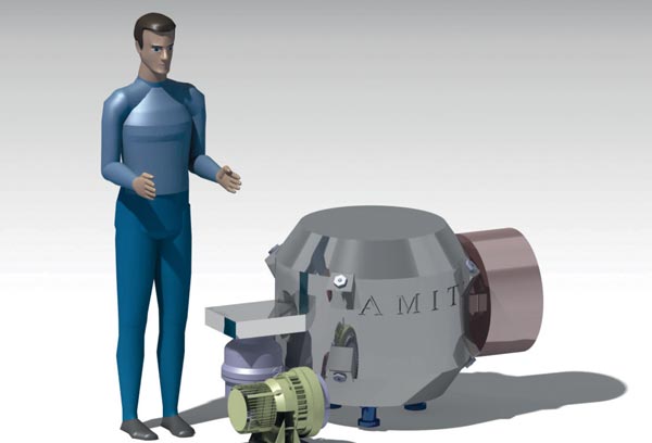
Image credit: CIEMAT.
Figure 5 shows a schematic of this cyclotron. A trade-off exists between increasing the magnetic field with higher levels of Lorentz stripping of negative ions and associated increases in the neutral beam-radiation field against the requirement of increasing the size of the radiation shielding around the cyclotron periphery. The nominal extraction radius for this machine will be around 11 cm. In other words, the size of the latest industrial medical-isotope producing cyclotrons have reverted to dimensions close to those of Lawrence’s first cyclotron developed over 80 years ago.


