High-energy physicists are proud that their instrumentation, which was developed for their research, has widespread applications in other fields. A recent symposium Special Applications of Particle Detectors in Medicine, Biology and Astrophysics (SAMBA), proved that this was not just wishful thinking.
The meeting, in Siegen, Germany, on 68 October, was highly cross-disciplinary, to the extent that it was sponsored by the Siegen Research Institute for Social Science and Humanities. The meeting highlighted the contribution of applied physics to our culture and to social prosperity.
After the formal opening by Siegen mayor Ulf Stötzel, Amos Breskin (Weizmann Institute, Israel) surveyed novel, large-area gas avalanche imaging photomultipliers for applications in the assessment of radiation damage to DNA, digital mammography and in the early detection of cancer. These aspects were underlined by Fulvia Arfelli (Trieste) who reported on the demanding diagnostic problems faced to reveal microcalcifications as possible indicators of breast cancer.
The prospects for macromolecular crystallography, particularly for proteins, using synchrotron facilities were presented by Peter Laggner (Graz, Austria). The development of multiwavelength anomalous dispersion techniques has transformed the method from an academic specialist’s playground into a large-scale, high-throughput industry. The importance of time-resolved measurements in X-ray structure analysis was emphasized for small- and wide-angle diffractive scattering experiments on non-crystalline materials.
The jumprelaxation technique opens up time domains down to microseconds, where supramolecular structure processes, induced by a change of parameters such as pressure, temperature or chemical environment, can be analysed. This kind of experiment calls for the development of new, fast, high-resolution and high-rate detectors so that the brilliance of the powerful synchrotron radiation sources, today and in the near future, can be exploited fully. With such experimental facilities designed as multipurpose, multi-user instruments, requirements emerge for new integrated approaches to measurement and detector control, fast data compactification, and artificial intelligence in the evaluation of multiframe results.
The domain of nuclear medicine is a good example of how difficult it is to replace cheap and reliable technology.
Commercial imaging systems for medical applications were reviewed by Detlef Mattern (Siemens/Erlangen). Despite ongoing attempts to switch to directly converting detectors, scintillators are still widely used for medical imaging. In radiography, computer tomography and nuclear medicine, a variety of scintillating devices are the workhorses in today’s clinical practice.
For radiography, flat X-ray detectors with evaporated scintillation layers are not far away. However, X-ray image intensifier tubes are competitive, and have features like speed and dynamic range that are still hard to beat. Although X-ray image intensifier tubes have disadvantages like size and weight, they give more robust image quality and have become cheaper over the decades. Detectors in computer tomography have evolved from scintillators into gaseous direct converters and back to scintillators again. Extreme timing requirements and detector modularity have ruled out designs that would rank as high performance in other fields.
The domain of nuclear medicine is a good example of how difficult it is to replace cheap and reliable technology. For many years, direct converters like cadmium telluride have been proposed as replacements for NaI(Tl) scintillation counters. However, for cost it is difficult to beat this light, high-output scintillator when used in combination with the practically noiseless photomultiplier. New ultracompact multianode photomultiplier designs could even make gamma cameras more compact without using directly converting detectors.
A highlight of the meeting was the talk by Gerhard Kraft (GSI Darmstadt, Germany) on the instrumentation for tumour therapy, which uses heavy-ion beams. Taking advantage of the Bragg peak at the end of the range of charged particles and the nonlinear biological sensitivity to the energy deposit, the small difference in destructive efficiency between healthy cells and cancer cells becomes sufficient to destroy well localized tumours accurately. Short-lived beta emitters, formed by carbon-12 beam fragmentation, are used to monitor the results via PET imaging.
High-granularity pixel counters, such as those being prepared for experiments at CERN’s LHC, are also used as focal detectors for X-ray astronomy. Lothar Strüder (Max Planck Institute for Extraterrestrial Physics, Munich) described the results of the ROSAT and Chandra missions, where radiation damage of semiconductor counters, caused by the Earth’s radiation belts, has to be taken into account. Similar problems concerning radiation hardness also have to be mastered for LHC detectors.
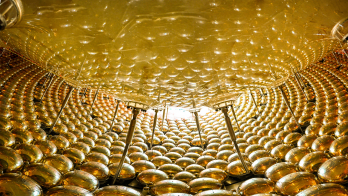
Allan Hallgren (Uppsala) concentrated on particle detectors for astrophysics. He described the virtues of neutrino astronomy using the Antarctic ice as Cherenkov medium. The search for dark matter, using cryogenic detectors, was covered by Klaus Pretzl (Bern). These cryogenic systems are also promising candidates for a new generation of particle trackers to be used in future accelerator experiments.
A public lecture by Claus Grupen (Siegen) entitled “Applications of particle detectors in astrophysics” attracted a good audience.
The meeting was organized jointly by H J Besch, C Grupen, N A Pavel, A Taune and A H Walenta. The proceedings will be published in a special volume of Nuclear Instruments and Methods.
Participants requested a follow-up meeting in this interdisciplinary field of detector applications.


